Home
/ Arteries Diagram ~ 0814 Diagram Of Artery And Vein Medical Images For Powerpoint Template Presentation Sample Of Ppt Presentation Presentation Background Images
Arteries Diagram ~ 0814 Diagram Of Artery And Vein Medical Images For Powerpoint Template Presentation Sample Of Ppt Presentation Presentation Background Images
Arteries Diagram ~ 0814 Diagram Of Artery And Vein Medical Images For Powerpoint Template Presentation Sample Of Ppt Presentation Presentation Background Images. As with veins, arteries are comprised of three layers: It is returned to the heart in the veins. In this image, you will find external carotid artery, internal carotid artery, vertebral artery, aorta and arch, pulmonary artery, cardiac artery, thoracic aorta, celiac trunk, superior mesenteric artery, renal artery, gonadal artery, inferior mesenteric artery, common iliac artery, external iliac artery. Spinal cord and meninges 12p image quiz. When there is a heart artery blockage, blood supply to areas of the heart are affected.
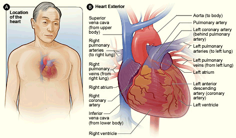
Arteries carry blood away from the heart in two distinct pathways: Immediately distal to the teres major, the brachial artery gives rise to the profunda brachii (deep artery), which travels with the radial nerve in the radial groove of the humerus and supplies structures in the posterior. There are three arteries that run over the surface of the heart and supply it with blood (see the diagram above). Coronary arteries supply oxygenated blood to the heart muscle, and cardiac veins drain away the blood once it has been deoxygenated. It is the main supply of blood for the arm.
1 from Immediately distal to the teres major, the brachial artery gives rise to the profunda brachii (deep artery), which travels with the radial nerve in the radial groove of the humerus and supplies structures in the posterior. The heart receives its own supply of blood from the coronary arteries. The one on the right is known as the right coronary. Ear anatomy 17p image quiz. Because the rest of the body, and most especially the brain, needs a steady supply of oxygenated blood that is free of all but the slightest. The cardiovascular system consists of the heart, blood vessels, and the approximately 5 liters of blood that the blood vessels transport. Two major coronary arteries branch off from the aorta near the point where the aorta and the left ventricle meet. Anterior view of the upper extremity 16p image quiz.
Each artery is a muscular tube lined by smooth tissue and has three layers:
Anterior view of the upper extremity 16p image quiz. After receiving blood directly from the left ventricle of the heart, the. On the left side, which is the main side, we have the left anterior descending (lad) that runs down the. It is the main supply of blood for the arm. It is a central communication that unites the internal carotid and vertebrobasilar systems. Classifica'on*of*arteries* • elas'c*arteries* *(conduc'ng*arteries) *aorta,*brachiocephalic,* commoncarod,* subclavian, vertebral,pulmonary,common* iliac. Heart posterior 20p image quiz. The one on the right is known as the right coronary. Because the rest of the body, and most especially the brain, needs a steady supply of oxygenated blood that is free of all but the slightest. The heart receives its own supply of blood from the coronary arteries. The typical configuration consists of two coronary arteries, a left main coronary artery (lmca) and a right coronary artery (rca), arising from the left posterior and right anterior aortic or coronary sinuses respectively, in the proximal ascending aorta.these are the only two branches of the ascending aorta. By definition, an artery is a vessel that conducts blood from the heart to the periphery. John bavosi/science photo library/getty images.
The one on the right is known as the right coronary. Lower limb arteries 17p image quiz. Anatomy and function of the coronary arteries. Arteries and veins are two of the body's main type of blood vessels. Two major coronary arteries branch off from the aorta near the point where the aorta and the left ventricle meet.
3 Anatomy Of The Coronary Arteries Atrain Education from www.atrainceu.com Learn vocabulary, terms, and more with flashcards, games, and other study tools. The typical configuration consists of two coronary arteries, a left main coronary artery (lmca) and a right coronary artery (rca), arising from the left posterior and right anterior aortic or coronary sinuses respectively, in the proximal ascending aorta.these are the only two branches of the ascending aorta. The tunicae intima, media, and externa. Simplified diagram of the human arterial system in anterior view. The right and left subclavian arteries give rise to the thyrocervical trunk. It is returned to the heart in the veins. The superior vena cava is the large. The heart receives its own supply of blood from the coronary arteries.
Two major coronary arteries branch off from the aorta near the point where the aorta and the left ventricle meet.
There is a point at which the anterior and posterior arterial circuits of the brain unite or anastomose. Classifica'on*of*arteries* • elas'c*arteries* *(conduc'ng*arteries) *aorta,*brachiocephalic,* commoncarod,* subclavian, vertebral,pulmonary,common* iliac. Proximal structures of the upper extremity 7p image quiz. Because the rest of the body, and most especially the brain, needs a steady supply of oxygenated blood that is free of all but the slightest. The superior vena cava is the large. Original vintage human anatomy victorian bookplate print 1890s medical diagram veins arteries blood circulatory system of the human body thepapermuseum. Arteries are components of the cardiovascular system. The neck is supplied by arteries other than the carotids. Learn the differences between an artery and a vein. An artery is an elastic blood vessel that transports blood away from the heart. The right coronary artery courses in the right atrioventricular groove. Simplified diagram of the human arterial system in anterior view. Over the years, cholesterol plaques can narrow the arteries supplying blood to the heart.
By definition, an artery is a vessel that conducts blood from the heart to the periphery. The cardiovascular system consists of the heart, blood vessels, and the approximately 5 liters of blood that the blood vessels transport. Coronary arteries supply oxygenated blood to the heart muscle, and cardiac veins drain away the blood once it has been deoxygenated. The superior vena cava is the large. The anterior tibial artery forms the arcuate artery and its many branches to supply blood to the top of the foot.
Gen Anatomy Major Arteries Diagram Diagram Quizlet from o.quizlet.com The one on the right is known as the right coronary. Human body artery diagram in detail. Blood is pumped from the heart in the arteries. An artery is an elastic blood vessel that transports blood away from the heart. Proximal structures of the upper extremity 7p image quiz. There are three arteries that run over the surface of the heart and supply it with blood (see the diagram above). It is the main supply of blood for the arm. The superior vena cava is the large.
Arteries are components of the cardiovascular system.
Original vintage human anatomy victorian bookplate print 1890s medical diagram veins arteries blood circulatory system of the human body thepapermuseum. It is returned to the heart in the veins. Each of these arteries delivers blood to the leg and continues into the foot, with the posterior tibial and fibular arteries forming the plantar arteries and plantar arch that supply blood to the bottom of the foot and toes. There are three arteries that run over the surface of the heart and supply it with blood (see the diagram above). On the left side, which is the main side, we have the left anterior descending (lad) that runs down the. The right coronary artery courses in the right atrioventricular groove. After receiving blood directly from the left ventricle of the heart, the. Coronary arteries supply blood to the heart muscle. The typical configuration consists of two coronary arteries, a left main coronary artery (lmca) and a right coronary artery (rca), arising from the left posterior and right anterior aortic or coronary sinuses respectively, in the proximal ascending aorta.these are the only two branches of the ascending aorta. There is a point at which the anterior and posterior arterial circuits of the brain unite or anastomose. The neck is supplied by arteries other than the carotids. Arteries are components of the cardiovascular system. Heart bypasses are either arteries or veins taken from other parts of the.
Share :
Post a Comment
for "Arteries Diagram ~ 0814 Diagram Of Artery And Vein Medical Images For Powerpoint Template Presentation Sample Of Ppt Presentation Presentation Background Images"


Post a Comment for "Arteries Diagram ~ 0814 Diagram Of Artery And Vein Medical Images For Powerpoint Template Presentation Sample Of Ppt Presentation Presentation Background Images"